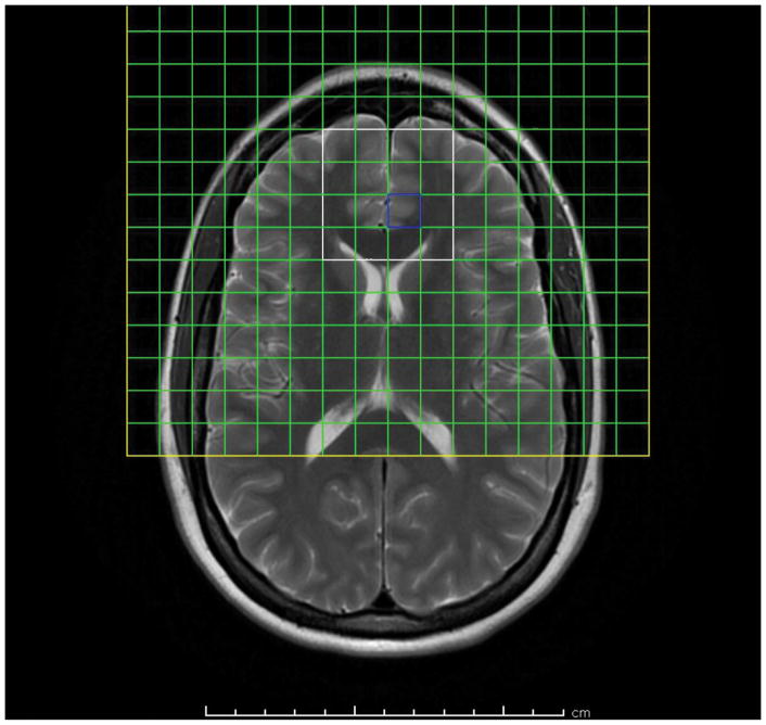
Figure 1.
T2-weighted axial-oblique (parallel to genu-splenium line) MRI of the human brain showing prescription of anterior cingulate 1H MRSI slab (yellow box). Usable spectra are obtained from the voxels (green squares) within the PRESS excitation volume (white box). Posterior mesial voxels within the slab sample left and right pregenual anterior cingulate cortex (LpAC, RpAC), anterior mesial voxels sample mesial superior frontal cortex, lateral voxels sample prefrontal white matter (anterior corona radiata).

