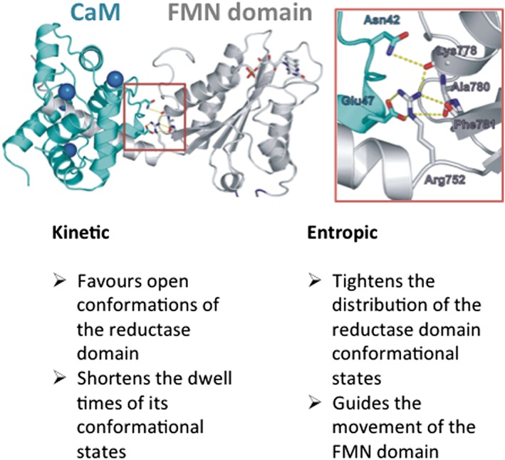
Figure 3.

CaM binding and effect on the conformational behaviours of the NOS reductase domain. The picture shows CaM (blue‐green) bound to an FMN domain‐CaM site construct of iNOS (grey) (Xia et al., 2009), with bound calcium ions coloured blue. The boxed area, magnified to the right, illustrates a key stabilizing interaction that involves a conserved Arg residue of the FMN domain (Arg752 of rat nNOS shown), and other residues as indicated, with CaM residue Glu47, that is required for the effects of CaM on NOS. The lower portion of the figure lists some kinetic and entropic effects of CaM binding. See text for related details.
