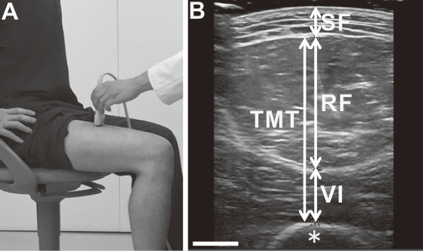
Fig. 1.
Measurement of thigh muscles using ultrasound.
Fig. 1a: Position of participant and ultrasound probe. The linear probe was set at midpoint of the right thigh in the sitting position.
Fig. 1b: Representative image of ultrasound. Thigh muscle thickness (TMT) was defined as the distance between the anterior fascia of rectus femoris muscle (RF) and posterior fascia of vastus intermedius muscle (VI) at the axial image. SF, subcutaneous fat; asterisk, femoral bone. Scale bar = 10 mm.

