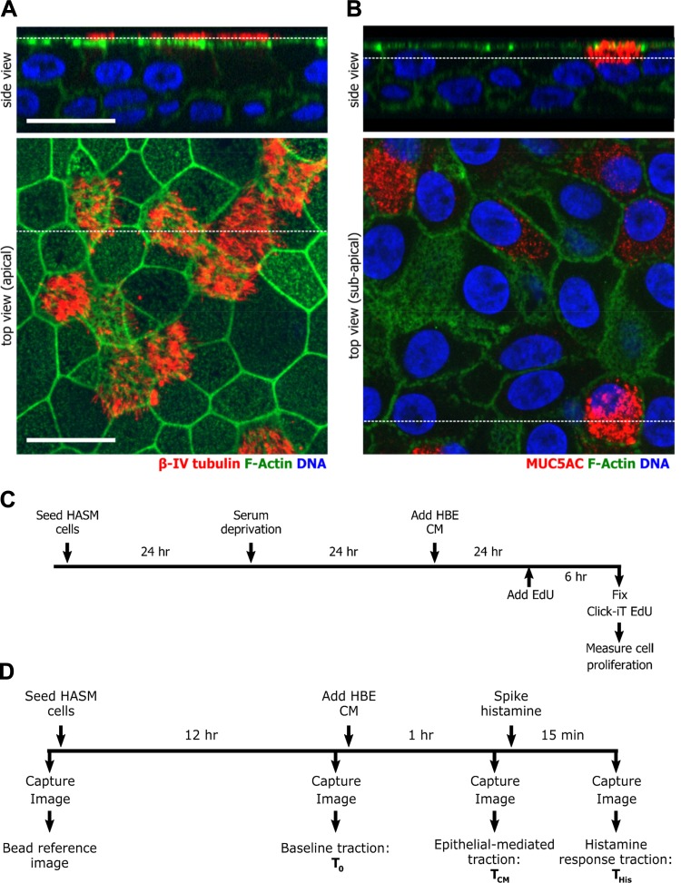
Fig. 1.
Experimental schema. A and B: primary human bronchial epithelial cells maintained in air-liquid interface culture were used for experiments when cells were well differentiated as determined by staining for βIV-tubulin (A), a ciliated cell marker, and for MUC5AC (B), a goblet cell marker. Cells were costained for F-actin and DNA (Hoechst). Broken white lines indicate the location in the corresponding image of the orthogonal cross section. The scale bar is 20 μm. C and D: timeline of the experimental procedures to measure proliferation (C) or contraction (D) of human airway smooth muscle (HASM) cells.

