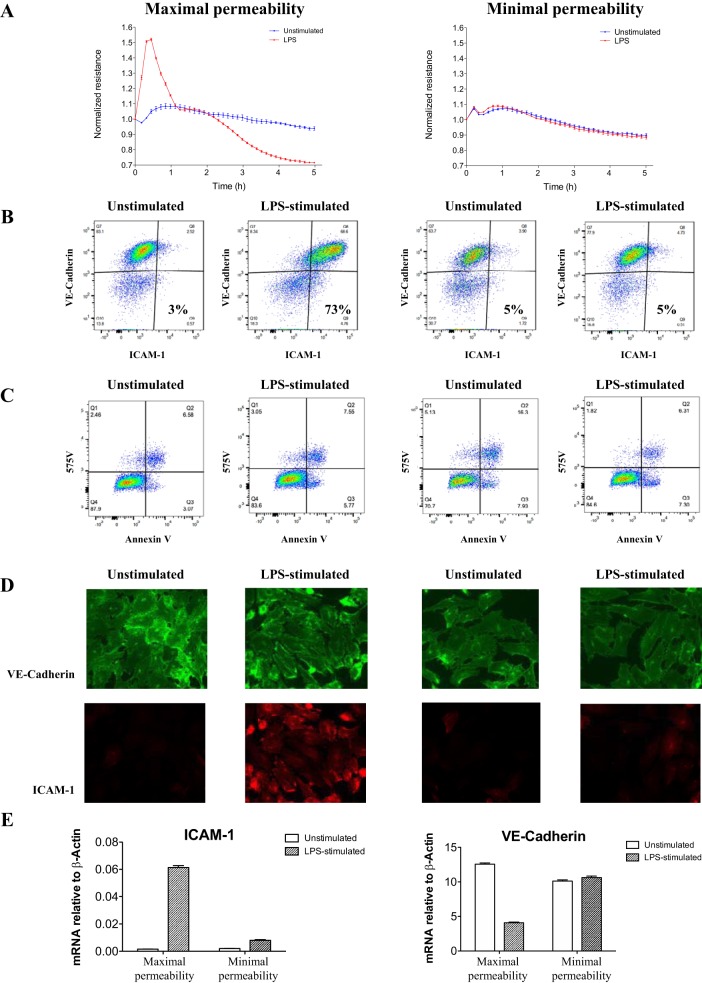
Fig. 3.
Variation in pulmonary endothelial cell intercellular adhesion molecule (ICAM)-1 and vascular endothelial (VE)-cadherin protein and gene expression after a 5-h exposure to leukocyte culture supernatants derived from representative patients with early sepsis in the maximal and minimal permeability group. A: endothelial cell resistance over 5 h of continuous electric cell-substrate impedance sensing (ECIS) measurement. B and C: flow cytometry scatter plots of ICAM-1 and VE-cadherin expression (B) and viability (575V) and apoptosis (annexin V) (C). D: immunohistochemistry of ICAM-1 and VE-cadherin expression. E: quantitative PCR of ICAM-1 and VE-cadherin gene expression showing an increase in ICAM-1 and decrease in VE-cadherin only in the maximal response group. Error bars represent SE of 3 technical replicates.

