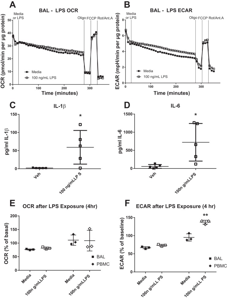
Fig. 6.
Acute lipopolysaccharide (LPS) challenge does not alter bronchoalveolar lavage (BAL) macrophage (MAC) bioenergetics. BAL MAC were subjected to extracellular flux analyses in which the oxygen consumption rate (OCR; A) and extracellular acidification rate (ECAR; B) were measured in response to exposure of 100 ng/ml LPS for 4 h were then of the cells. After 4 h of exposure, a mitochondrial stress test was performed using sequential addition of site-specific inhibitors of the electron transport chain [oligomycin (Oligo), FCCP, and rotenone/antimycin A (Rot/Ant. A)]. A representative plot is shown, n = 6. OCR and ECAR values are expressed normalized for protein content. Levels of IL-1β (C) and IL-6 (D) produced by BAL MAC after 4 h challenge with media or 100 ng/ml LPS, n = 5. BAL MAC and peripheral blood monocytes (PBMC; E and F) from the same subject were stimulated with media or 100 ng/ml LPS for 4 h before measurement of their OCR and ECAR. Data are expressed as a percent of the baseline value. *P < 0.05 from vehicle (Veh) or media controls, means ± SD, n ≥ 3.

