Fig. 4.
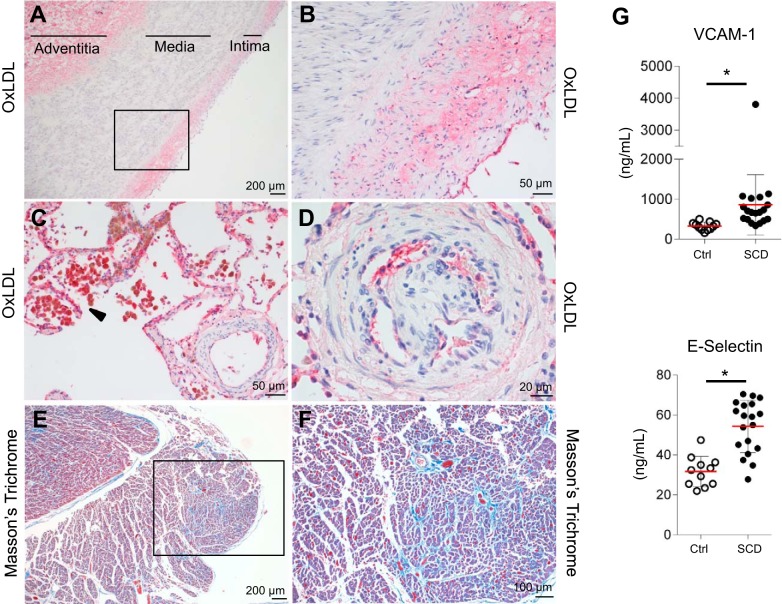
Vascular injury localized with lipid oxidation is observed in the pulmonary artery, lung tissue, and right heart ventricle of deceased patients with SCD. This coincided with observations of endothelial dysfunction markers in plasma of SCD and control patients. Vascular injury in pulmonary artery, lung tissue, and right heart ventricle of deceased patients with SCD and endothelial dysfunction markers in plasma of SCD to control group. A and B: thickened intimal and medial compartments of the pulmonary artery of deceased patients with SCD showed positive staining for oxLDL in the enlarged intimal layer and adventitia but not in the thickened media compartment. C: macrophage accumulation around lung vessels with intensified staining for oxLDL. D: endothelium of precapillary vessels stained positive for oxLDL and showed unregulated endothelial proliferation. E and F: right heart ventricle fibrosis was seen in MT staining. G: endothelial dysfunction markers such as soluble VCAM-1 and E-Selectin were significantly elevated in plasma of SCD over Ctrl. *P < 0.05, significant difference compared with Ctrl group. Ctrl, control; oxLDL, oxidized LDL; MT, Masson’s trichrome; SCD, sickle cell disease.

