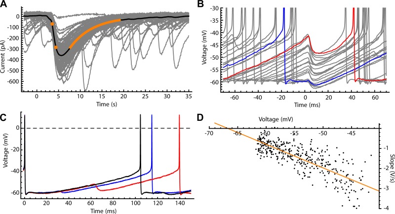
Fig. 2.
Synaptic inhibition of substantia nigra pars reticulata (SNr) neurons by direct pathway axons. A: photo-evoked direct pathway inhibitory postsynaptic currents (IPSCs) recorded from SNr neurons in whole cell configuration using CsCl intracellular solution (gray traces). Mean photo-evoked IPSC is shown in black, illustrating the measurement of rise time (20–80%, orange dots) and decay time constant (from the illustrated single-exponential fit, orange line). B: sample of photo-evoked inhibitory postsynaptic potentials (IPSPs) recorded in perforated-patch configuration (gray). IPSP amplitude is sensitive to membrane voltage at the time of onset; IPSPs earlier in the interspike interval (ISI) (e.g., blue) occur at more negative membrane potentials and are smaller than IPSPs later in the ISI (red). C: ISI voltage trajectories with no IPSP (black), an early IPSP (blue), and a late IPSP (red). Both early and late IPSPs lengthen the ISI, but late IPSPs have a larger effect. D: chloride reversal potential measured in the perforated-patch configuration. The IPSP onset slope was plotted vs. the membrane potential. A linear fit (orange) was obtained by least-squares linear regression, and the x-intercept of the fit line (where IPSP slope = 0) was taken as reversal potential (Erev).

