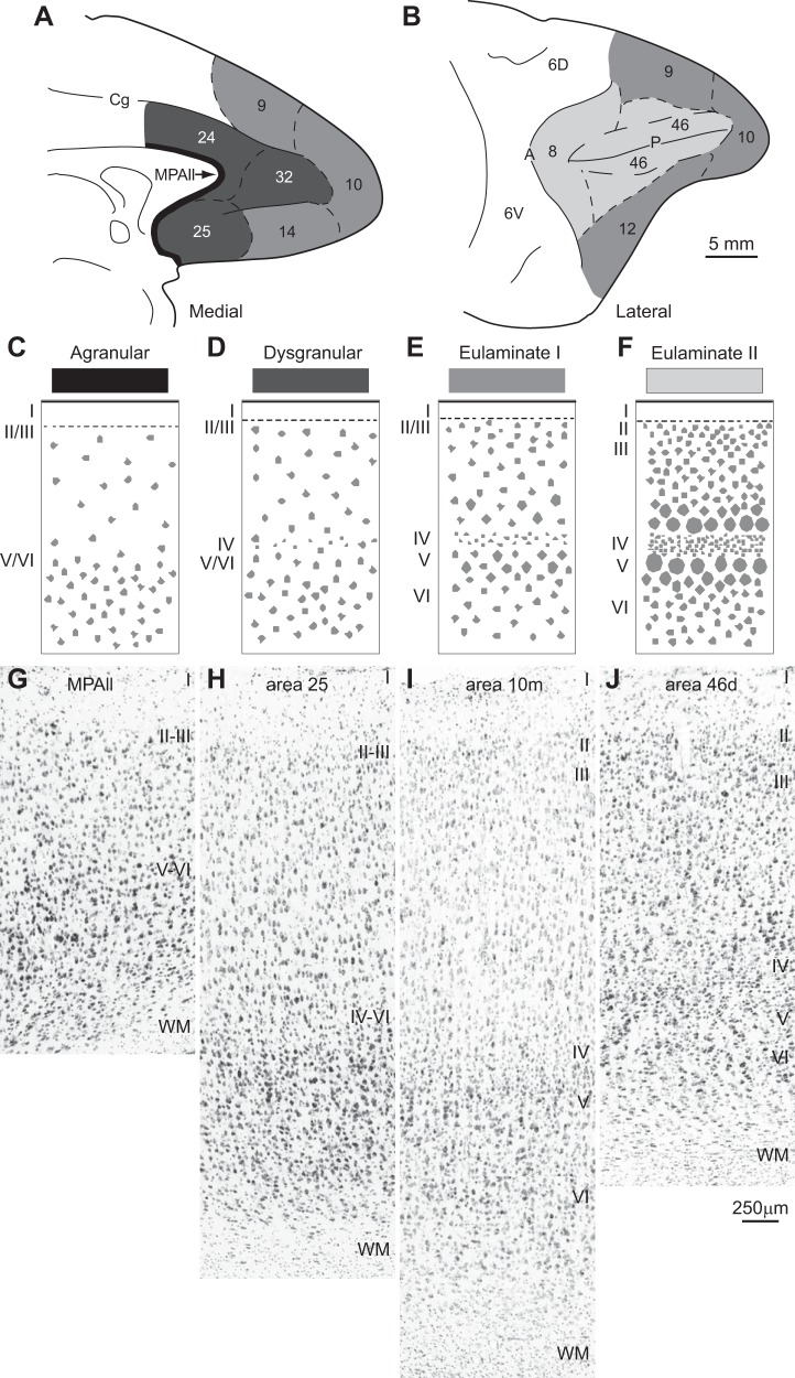
Fig. 1.
Systematic variation in cortical laminar structure using the prefrontal cortical region as a model system. Laminar structure is used to group areas into types of cortex, indicated by gray shading. The number of types depicted may vary depending on the region (system) and the desired level of resolution. A: medial view of the frontal lobe shows areas with the lowest (agranular; black) through areas with increasing elaboration of laminar structure (shown by lighter shades of gray). B: lateral view of the frontal lobe shows prefrontal areas with the greatest laminar elaboration (lightest gray). C–F: cartoons depict different types of cortex. C: agranular cortex has no layer IV. D: dysgranular cortex has an incipient layer IV. E: eulaminate area with 6 layers and a moderately dense layer IV. F: eulaminate area with a well-developed layer IV and better distinction between other layers. G–J: photomicrographs of coronal sections taken through different prefrontal areas. G: agranular medial prefrontal area MPAll (medial periallocortex). H: the dysgranular part of area 25. I: eulaminate I area 10m. J: eulaminate II area 46d. WM, white matter. Arabic numerals indicate prefrontal areas according to Barbas and Pandya (1989). Roman numerals indicate cortical layers. Calibration bar in J applies to G–J.

