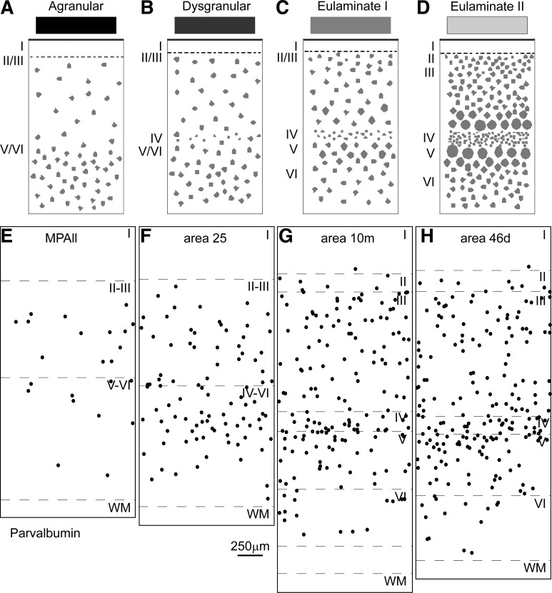
Fig. 5.
Systematic variation in the laminar structure of the cortex is accompanied by other features. A–D: cartoons show systematic changes in laminar structure that can be used to group areas into types of cortex. Shades of gray (top) show types of cortices from the least (black) to the greatest (lightest gray) elaboration of laminar structure. E–H: the laminar structure of the cortex is accompanied by other cellular and molecular features. In the prefrontal cortex, the trend is seen in the distribution of the neurochemical class of parvalbumin (PV) inhibitory neurons, which show the lowest density in agranular areas (E) and increasing density through sequential types of areas with increasing elaboration of laminar structure (F–H). E: agranular medial prefrontal area MPAll (medial periallocortex). F: the dysgranular part of area 25. G: eulaminate I area 10m. H: eulaminate II area 46d. WM, white matter. Roman numerals indicate cortical layers. Calibration bar in F applies to E–H.

