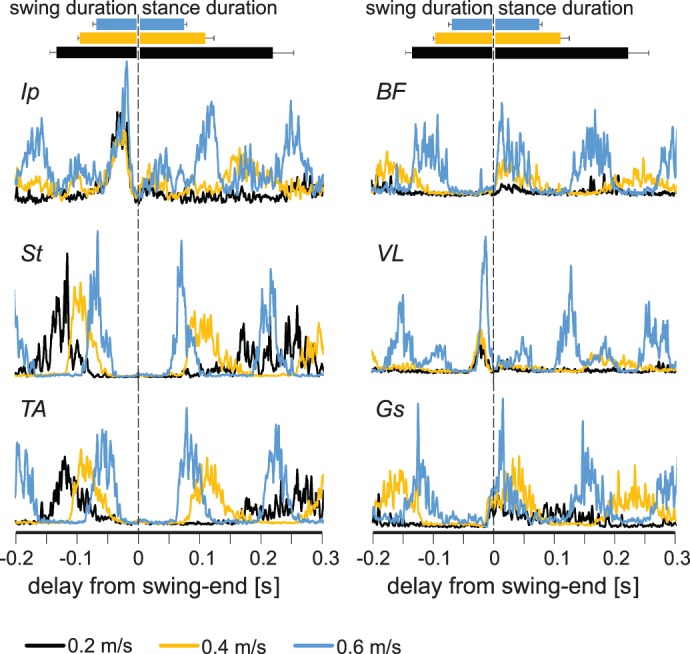
Fig. 4.

Electromyogram (EMG) activity in leg muscles increases at higher walking speeds. Average EMG activities (rectified and smoothed) from all flexor (left) and extensor (right) muscles at different speeds (black, 0.2 m/s; yellow, 0.4 m/s; blue, 0.6 m/s) recorded in this study indicate that the EMG activity of extensor muscles increases at higher speeds. Durations of stance and swing are shown by horizontal bars (top) indicating averages ± SD. Ip, iliopsoas; BF, anterior biceps femoris; St, semitendinosus; VL, vastus lateralis; TA, tibialis anterior; Gs, gastrocnemius.
