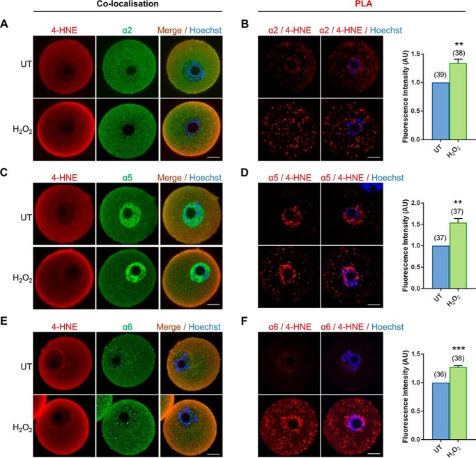
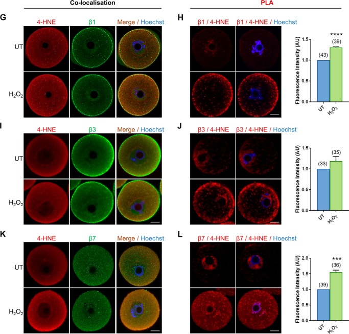
Figure 5.
4-HNE adducts to multiple subunits of the core proteasome. GV oocytes were treated with 35 μm H2O2 (H2O2) for 1 h to induce oxidative stress and were prepared for immunofluorescence analysis and proximity ligation assays alongside their untreated (UT) counterparts to examine the susceptibility of proteasome subunits to 4-HNE modification. A, C, E, G, I, and K, for immunofluorescence analysis, all proteasome subunits examined (α2, α5, and α6 and β1, β3, and β7) were detected in the oocyte and co-localized with 4-HNE, particularly at the periphery of the oocyte after H2O2 treatment using appropriate anti-proteasome (green) and anti-4-HNE (red) antibodies, and nuclei were counterstained with Hoechst (blue). B, D, F, H, J, and L, PLA between 4-HNE and several proteasome subunits (α2, α5, and α6 and β1, β3, and β7) revealed their susceptibility to 4-HNE modification with fluorescent foci (red) evident throughout the oocyte that significantly intensified after oxidative insult, with the exception of β3. Nuclei were counterstained with Hoechst (blue). Scale bar = 20 μm. These experiments were repeated across three independent biological replicates, with a minimum of 10 oocytes, pooled from a minimum of three animals. Statistical analyses were performed using Student's t test, **, p ≤ 0.01; ***, p ≤ 0.001; and **** p ≤ 0.0001. Data are presented as mean of three replicates ± S.E. AU, arbitrary units.


