
Figure 2.
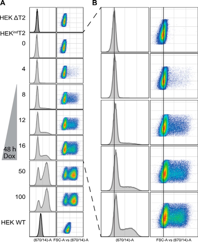
Flow cytometric quantification of induced GalNAc-T2. A, cells were induced for 48 h with doxycycline (Dox) before being fixed, permeabilized, and stained with anti-GalNAc-T2 primary antibody and Alexa Fluor 647 secondary antibody. B, enlargement of HEKindT2 induced from 0 to 16 ng/ml doxycycline with a help line centered at the peak of 0 ng/ml. The induction is biphasic with a distinct global right shift of the lower-expressing population. Data are representative of two biological and two technical replicates.