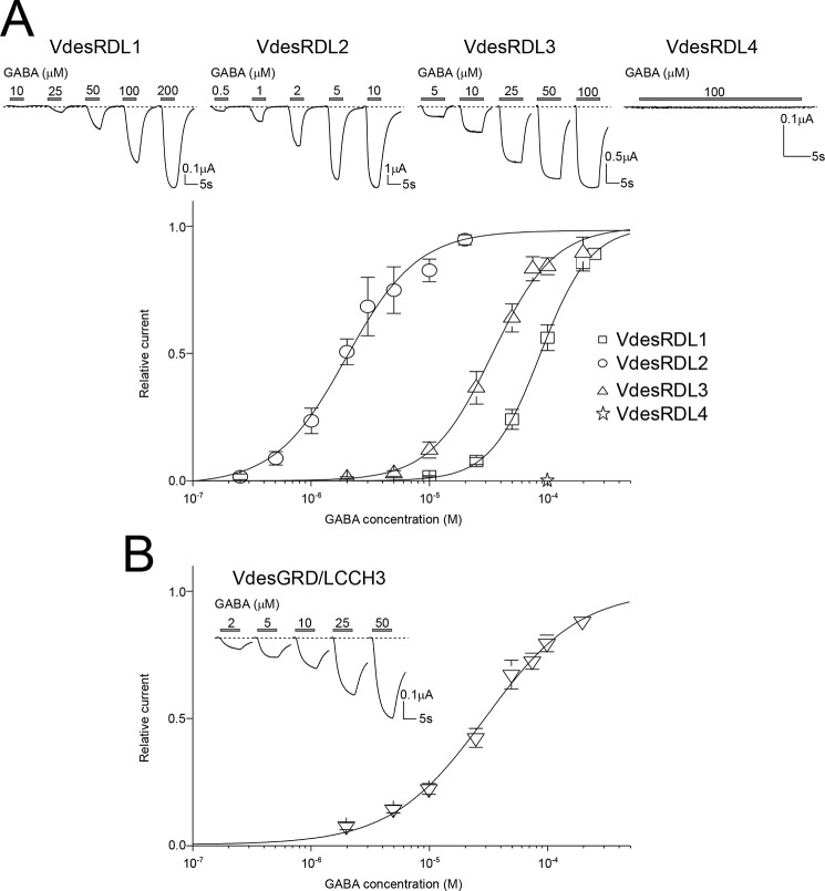
Figure 2.
GABA-evoked currents for VdesRDLs and VdesGRD/LCCH3 expressed in X. laevis oocytes. A, top panel, representative current traces obtained with increasing GABA concentrations in X. laevis oocytes after the injection of the cRNAs encoding VdesRDL1, VdesRDL2, VdesRDL3, or VdesRDL4. The duration of GABA perfusion was adjusted for each oocyte to reach the maximal current amplitude. Note the absence of current in oocytes injected with the cRNA encoding VdesRDL4. Bottom panel, GABA concentration-response curves obtained in X. laevis oocytes that express VdesRDL1, VdesRDL2, or VdesRDL3. B, same as in A but in X. laevis oocytes that express VdesGRD/LCCH3. The data are the means ± S.E. of n = 9–23 oocytes.

