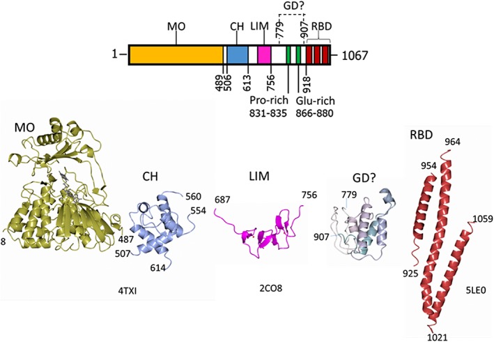
Figure 1.

Domain organization of human MICAL1 and structures of the isolated domains. The numbering indicates the boundaries of the known domains of MICAL1: MO, monooxygenase‐like FAD‐containing domain; CH, type 2 calponin homology domain; LIM, LIM domain containing two zinc finger motifs; GD? indicates a potential globular domain identified by Robetta modelling, which contains the Pro‐rich motif typical of regions binding to SH3 domains and a Glu‐rich region; RBD, Rab‐binding domain as identified by 45, 46. The experimental (4TXI, 2CO8, 5LE0) and modeled (GD?) structures of the MICAL1 domains are also shown in ribbon. These and other structural models have been drawn with CCP4 MG.70
