
Figure 3.
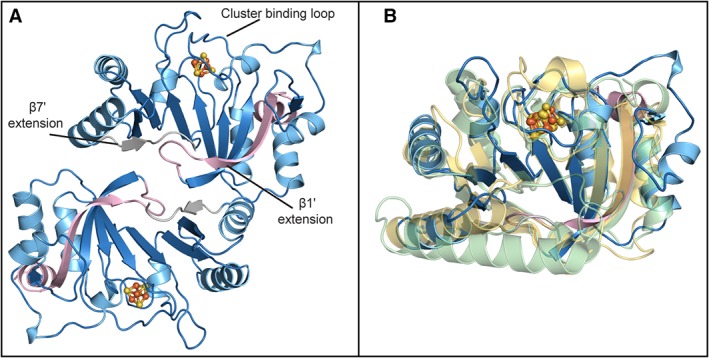
Structure of QueE from Escherichia coli. (A) Structure of EcQueE, shown as ribbons, folds into a head‐to‐tail functional dimer with the dimer interface composed of interactions between the N‐terminal (light pink) and C‐terminal (grey) extensions. The modified AdoMet core, a partial (β6/α5) TIM barrel, is shown in blue. (B) EcQueE (blue) monomer overlays well with the monomers of BsQueE, (PDB ID 5TH5), (translucent light green) and BmQueE, (PDB ID 4NJI), (translucent yellow). In both panels, [4Fe–4S] clusters are shown in a ball and stick representation, where iron is colored orange and sulfur is colored yellow.