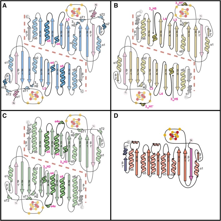
Figure 4.

Topology diagrams for QueE homologs and PFL‐AE. (A) EcQueE, (B) BmQueE, (C) BsQueE and (D) PFL‐AE. The core AdoMet domains are colored blue for EcQueE, yellow for BmQueE and green for BsQueE whereas the N‐ and C‐terminal extensions are colored light pink and grey (respectively) for all three QueE structures. The differences between the QueE homologs structures are shown in bold and the corresponding secondary structure element denoted in magenta and the dashed line delineates the QueE dimer interface. The topology diagram of PFL‐AE is shown with the N‐ and C‐terminal extensions colored pink and slate respectively and AdoMet domain colored in coral. The iron atoms of the [4Fe–4S] clusters are colored orange and sulfur atoms are colored yellow. Cysteine ligands to the [4Fe–4S] cluster are shown as yellow circles. Structural elements outside the AdoMet radical core fold are labeled with a prime.
