
Figure 5.
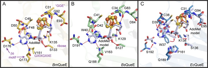
AdoMet binding pocket in QueE homologs. AdoMet binding within the AdoMet core (translucent ribbons) is shown for (A) BmQueE (PDB ID 4NJI), (B) BsQueE (PDB ID 5TH5) and (C) EcQueE. In (A), AdoMet binding motifs are labeled in magenta. See Fig. S3 for stereo views and further description of AdoMet binding. The binding pockets are composed of residues (sticks), which can provide hydrogen bonds (red) to AdoMet (white). The irons (orange) and the sulfurs (yellow) of the [4Fe‐4S] AdoMet radical cluster are shown as spheres. In (B), the intact AdoMet molecule is modeled using the adenosyl moiety of the 6‐carboxypterin‐5′‐deoxyadenosyl ester adduct (PDB ID 5TH5) and an intact AdoMet molecule (PDB ID 4NJI) as a guide. The AdoMet binding pocket of EcQueE (blue) is shown overlaid with the binding pocket from BmQueE (white) to highlight the changes that need to be made (red arrows) to allow binding of the modeled AdoMet (white) molecule.