Figure 8.

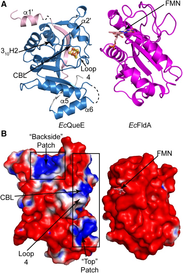
Electrostatic surface charge for EcQueE and the cognate Fld, EcfldA. (A) Ribbon drawing of monomer of EcQueE with the AdoMet radical core in blue and the N‐ and C‐terminals in light pink and grey, respectively, oriented such that the predicted binding sites are facing EcFldA. The structure of EcFldA (PDB ID 1AHN) is also shown in ribbon representation (magenta) with the FMN cofactor and the loops proposed to bind partner proteins facing EcQueE. (B) The solvent accessible electrostatic surface representations of EcQueE and EcFldA with FMN colored salmon are also displayed in the same orientation as in A. Electrostatic potentials are depicted on a colorimetric scale from red to blue for −1 to +1 kTe−1.
