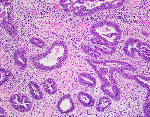
FIG. 2.

Serous carcinoma with a glandular pattern. Serous differentiation can be recognized at intermediate-power magnification by the presence of cells with high nuclear-to-cytoplasmic ratios and at high power by the presence of enlarged and atypical nuclei. Confirmatory endometrioid features are lacking. The diagnosis can be confirmed with a p53 immunohistochemical stain, which shows mutation-type staining in at least 90% of serous carcinomas. Although mutation-type p53 staining can be present in endometrioid carcinomas, almost all are International Federation of Gynecology and Obstetrics grade 3 carcinomas, which glandular serous carcinomas do not resemble.
