Fig. 3.
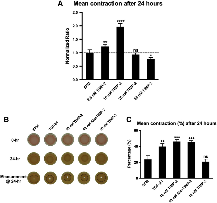
Fibroblast-mediated 3D collagen matrix remodeling. (A) Differential effect of various concentrations of TIMP-2 on 3D collagen ECM remodeling as assessed by extent of contraction over time: TIMP-2 stimulated collagen ECM contraction at lower concentrations (2.5 and 10 nM), whereas the highest concentration (50 nM) inhibited ECM contraction when compared to the SFM group. Data presented were obtained from three individual experiments, and all values were normalized to the corresponding SFM control group. Bars represent mean±S.D. (N=7 per group). *P<.05; **P<.01; **** P<.0001. (B) Representative photographs of cell–ECM constructs at 0 and 24 h by treatment group. (C) Percentage of ECM contraction (%) from the initial surface area 24 h after release. TGF-beta1 (10 ng/ml), TIMP-2 and Ala+TIMP-2 significantly stimulated collagen ECM contraction, whereas TIMP-3 did not alter ECM contraction as compared to the SFM group. Bars represent mean±S.D. (N=3 per group). ** P<.01; *** P<.001; ns, nonsignificant.

