Fig. 4.
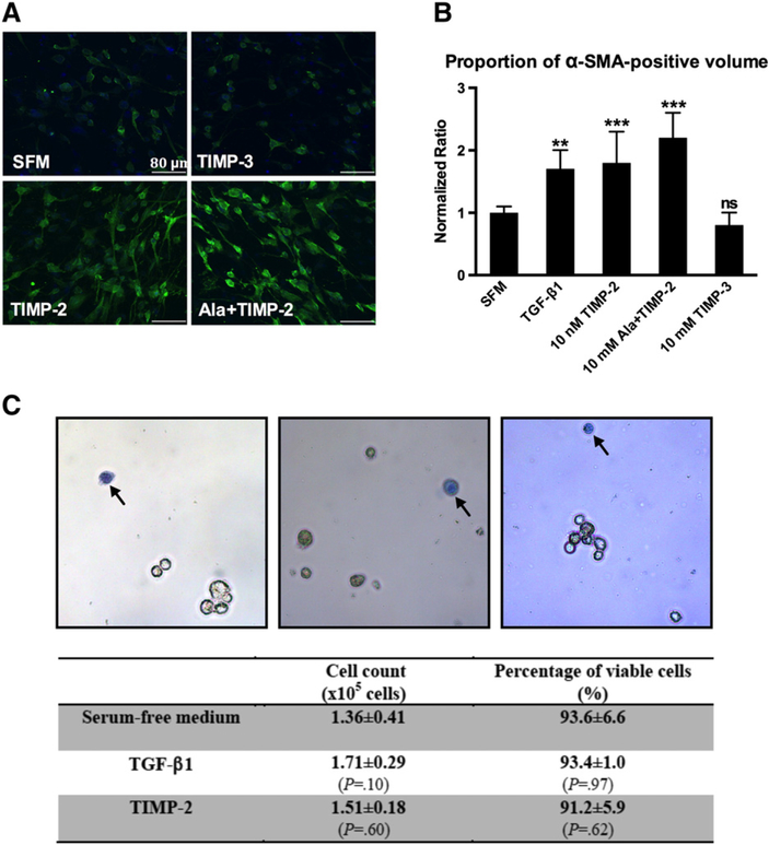
Cardiac myofibroblast activation and cell viability. (A) Representative confocal microscopic images of cardiac fibroblasts/myofibroblasts embedded in collagen ECM constructs from different treatment groups. Cells were stained for alpha-SMA (green) and for nuclei (DAPI, blue). Both TIMP-2 and Ala+TIMP-2 stimulated alpha-SMA expression and induced morphological transformation. TIMP-3 did not exert a similar effect. Scale bar=80 μm. (B) Proportion of confocal image volume stained positive for a-SMA (green). Values were normalized to that of the SFM group. TGF-beta1 (10 ng/ml), TIMP-2 and Ala-TIMP-2 induced a similar increase in expression of alpha-SMA. TIMP-3 did not alter alpha-SMA expression as compared to the SFM group. Bars represent mean±S.D. (N=8 random field images per group). **P<.01; *** P<.001; ns, nonsignificant. (C) Representative photomicrographs of cells harvested directly from cell–ECM constructs after the exposure to targeted treatments. Dead cells appear blue after trypan blue staining (arrows). Objective: 20×. All treatment groups yielded an equal cell number and cell viability (N=8 per group).

