Fig. 6.
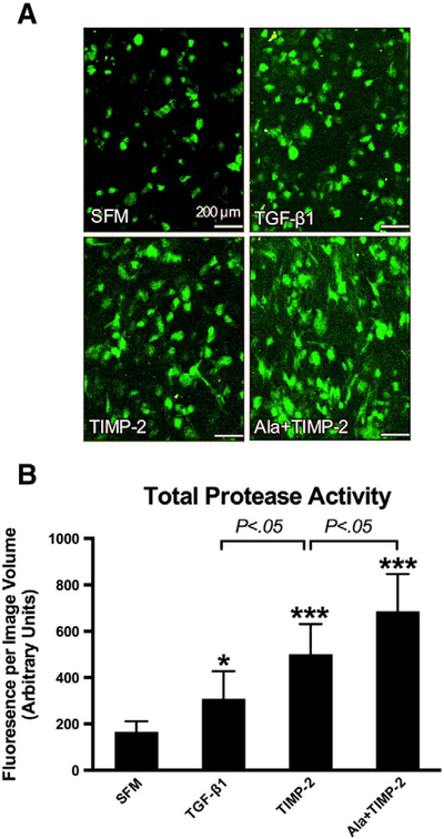
In situ zymography with quantitative assessments of total protease activity in collagen ECM microenvironment. (A) In situ zymography: representative confocal microscopic images of the emitted fluorescent signals (green) in collagen ECM, following proteolysis of the embedded DQ Gelatin-FITC. Scale bar=200 μm. (B) Total protease activity in cell–ECM constructs was quantified as total fluorescent signal per image volume. TGF-beta1 (10 ng/ml), TIMP-2 (10 nM) and Ala+TIMP-2 (10 nM) increased the total protease activity in the ECM microenvironment as compared to SFM. TIMP-2 yielded more protease activity than TGF-beta1 (P<.05). Ala+TIMP-2 resulted in a higher protease activity than TIMP-2, likely due to a lack of MMP inhibition (P<.05). Bars represent mean±S.D. (N=8 per group). * P<.05; *** P<.001 as compared to SFM.

