
Fig. 3.
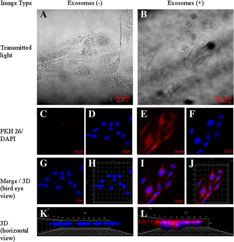
Cellular internalization of HUVEC-derived EVs into HUVECs. HUVECs were incubated for 24 h with EVs labelled with PKH26 (red). The carryover of PKH26 was observed when cells were incubated with PKH26 without EVs (negative control). a, b – Transmitted light. c, e – PKH26 staining. d, f – DAPI staining. g, i – Merged 2D view. h, j – Merged 3D view. k, l – 3D horizontal view