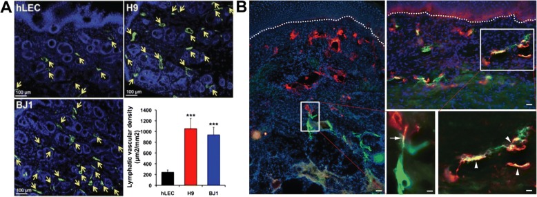
Fig. 4.
(a) LEC (LYVE-1+/Podoplanin+) cells derived from hPSCs (H9 and BJ1) were injected into the skin wound on the backs of nude mice. Lymphatic vessels indicated by arrows (LYVE-1) were significantly increased in mice injected with hPSC-LECs (H9 and BJ1) compared to the hLEC-control. ***p<0.001. Illustration in panel A was adapted with permission from [178]. (b) Fibrin/Collagen I hydrogels were used to generate dermo-epidermal skin grafts with blood and lymphatic capillaries. After 14 days post-transplantation, anastomosis occurred either as a “direct connection” (arrows) or as a “wrapping connection” (arrowheads). Dashed lines indicate the dermo-epidermal junction. Human lymphatic vessel (human podoplanin stained in red), rat lymphatic vessel (rat podoplanin stained in green), and nucleus stained in blue. Scale bars are 50 μm. Illustration in panel B was adapted with permission from [50]

