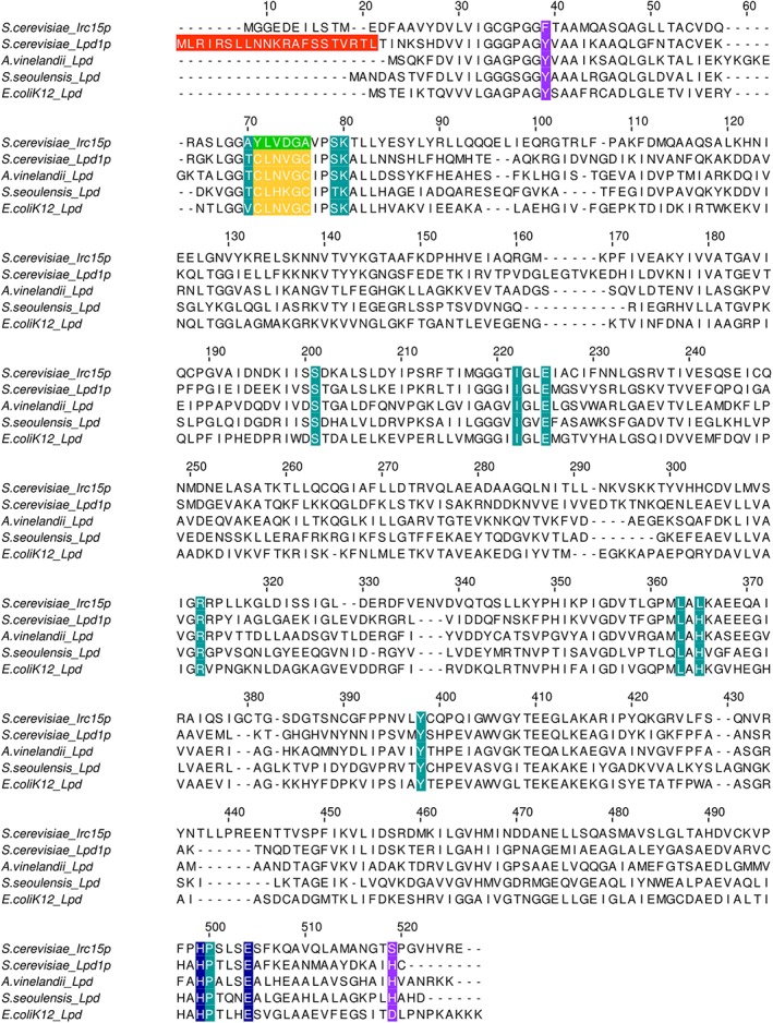
Figure 4.

Alignment of the Irc15p protein sequence with sequences of LPD from S. cerevisiae, E. coli, S. seoulensis and A. vinelandii. The mitochondrial targeting sequence of Lpd1p is highlighted in red. The amino acid signature near the redox‐active disulfide is highlighted in yellow. The respective sequence in Irc15p is highlighted in green. The catalytic His‐Glu diad is highlighted in blue. Other residues in the active site are highlighted in petrol. Residues involved in structural stabilization are highlighted in purple.
