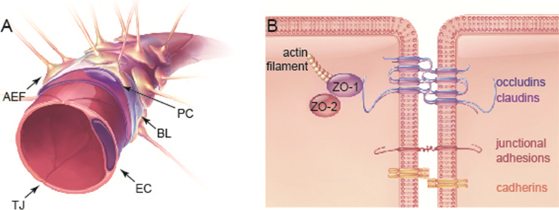
Fig.1.
The anatomy of the blood–brain barrier (BBB). (A) The BBB is comprised of astrocytic endfeet (AEF) and pericytes (PC), which surround a single layer of endothelial cells. Endothelial cells secrete extracellular matrix proteins, forming the basal lamina (BL). Endothelial cells are connected by tight junctions (TJ) that form a highly selective semi-permeable membrane, separating the brain’s parenchyma from the circulatory system. (B) TJs anchor endothelial cells close together through a number of transmembrane proteins (e.g. occludin, claudins and junctional adhesion molecules). These TJ proteins interact with cytoplasmic scaffolding proteins such as zonula occludens (ZO), as well as with the actin cytoskeleton. Other integral membrane proteins and cell adhesion molecules, such as cadherins and integrins, are also found at cell-cell junctions [105]. Exercise has been shown to increase the expression of claudins, as well as the co-expression of proteins from the ZO and occludin family at the plasma membrane.

