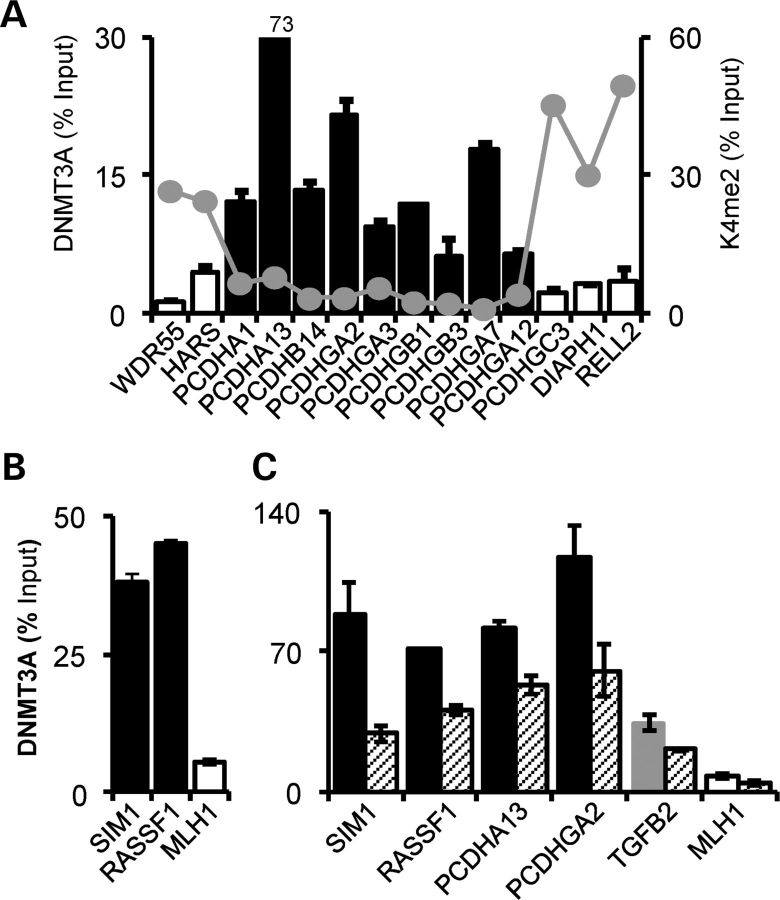
Figure 6.
Association of DNMT3A with tumour-specific methylation changes in Wilms' tumour WiT49 cells. (A) ChIP across the PCDH LRES on chromosome 5q31. High DNMT3A binding is apparent at hypermethylated genes (black bars) and lower binding at unmethylated genes (open bars). Histone 3 lysine 4 dimethylation (35) across the locus is shown by the grey points. (B) DNMT3A recruitment at non-LRES hypermethylated (SIM1 and RASSF1) and unmethylated (MLH1) genes. (C) WT1 knockdown leads to decreased DNMT3A recruitment at methylated genes. High DNMT3A is present at the hypermethylated genes (SIM1, RASSF1, PCDHA13 and PCDHGA2, black bars) and not at the unmethylated MLH1 (white bar). Note the intermediate level DNMT3A recruitment at TGFB2 (grey bar). DNMT3A levels after WT1 knockdown are indicated by hatched bars.

