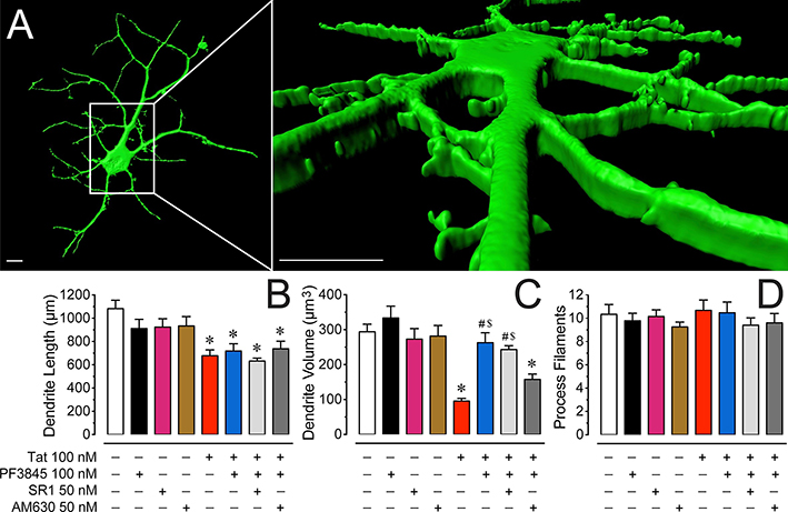
Figure 3. Morphological changes on dendrites of PFC neuronal cultures.
(A) Three-dimensional reconstruction of PFC neuronal dendrites at DIV 8 (B) Total dendritic length (μm) is significantly decreased by Tat 100 nM after 24 h. No protective effect was noted when pretreating Tat-exposed neurons with the FAAH enzyme inhibitor PF3845 100 nM. (C) In contrast, the significant reduction of total dendritic volume (μm3) by Tat 100 nM was prevented with pretreatment of PF3845 100 nM. Interestingly, whereas SR1 50 nM was not able to block the protective effects of PF3845 100 nM, the CB2R antagonist AM630 50 nM reduced dendritic volume comparably to the Tat 100 nM treatment condition. (D) The total number of process filaments were not significantly different between conditions. Statistical significance was assessed by ANOVAs followed by Bonferroni’s post hoc tests; *p < 0.01 vs. control, #p < 0.001 vs. Tat 100 nM, $p < 0.05 vs. AM630 50 nM + PF3845 100 nM + Tat 100 nM (at least three independent experiments). SR1: SR141716A. Scale bars = 10 μm.

