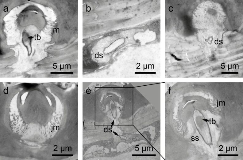
Figure 3.
Transmission electron microscopy images of setae on the hemelytra of N. glauca. a–d) Clavus. a) Tubular body (tb) at the base of the seta. Note the joint membrane (jm). b) Outer dendritic segment with the dendritic sheath (ds) sectioned below the cuticle. c) Part of the dendrite in the outer receptor lymph cavity of the sensillum. The base of the seta is visible above the dendrite. d) Base of the seta with the joint membrane (jm). e,f) Membranous part of the hemelytra. e) Dendrite with an apical tubular body attached to the base of the seta. Below more proximal parts of the dendrite are shown. f) Detail of e. The tubular body (tb) is visible. The socket septum (ss) is attached to the tip of the dendrite where the tubular body is located.

