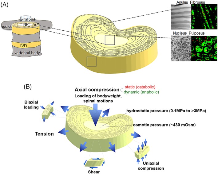
Figure 1.

(A) Schematic representation of spinal anatomy (left) and the heterogeneous tissue composition of the intervertebral disc (IVD) (right). Insets (right) show matrix organization (scanning electron microscopy, gray) and cell organization (fluorescence) for anulus fibrosus (AF) and nucleus pulposus (NP) regions. AF matrix is composed of aligned collagen I fiber bundles, while a much random distribution of collagen II bundles and proteoglycan is observed in the NP. Fluorescence insets are rat IVD cells stained for collagen VI (green) and nucleus (red) revealing the different cellular arrangement in each tissue area. Adapted from Cao et al174. (B) Mechanical deformation of the IVD. Axial compression can be either catabolic or anabolic depending on mode (static, dynamic), magnitude, frequency, and duration. Loading of the hydrated NP matrix results in hydrostatic and osmotic pressures. When the disc is axially loaded, the NP region becomes compressed and the AF undergoes radial and circumferential tension to limit overall disc expansion in the transverse plane. Adapted from Setton et al195
