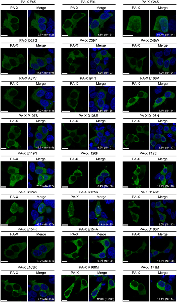
Figure 2. Intracellular localization of mutant PA-X.
293 cells were transfected with a plasmid encoding the mutant PA-X and fixed 24 h later. These cells were then stained with an anti-DYKDDDDK (FLAG) tag antibody (green). All images were obtained using confocal microscopy. The nuclei are stained with Hoechst 33342 (blue). The percentage of cells in which PA-X was localized both in the nucleus and the cytoplasm is indicated in each panel. The total number of counted cells is indicated in parentheses. Bars, 10 μm.

