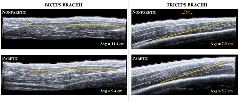
Figure 2.
Extended field-of-view ultrasound images of the long head of biceps brachii (left) and the distal portion of the lateral head of triceps brachii (right) in a single subject with chronic hemiplegia. Images were taken in the same neutral elbow posture for both muscles and in both the nonparetic (top) and paretic (bottom) limb. The dashed line in each image represent a single fascicle measurement as detailed in the methods, while the hash marks in each image represent 1 cm. The average values noted here were calculated over the 12 repeated measurements (3 images, 4 fascicle measurements each) in each arm and muscle for this participant. Schematic representation of muscle architecture can be viewed in previous work.33

