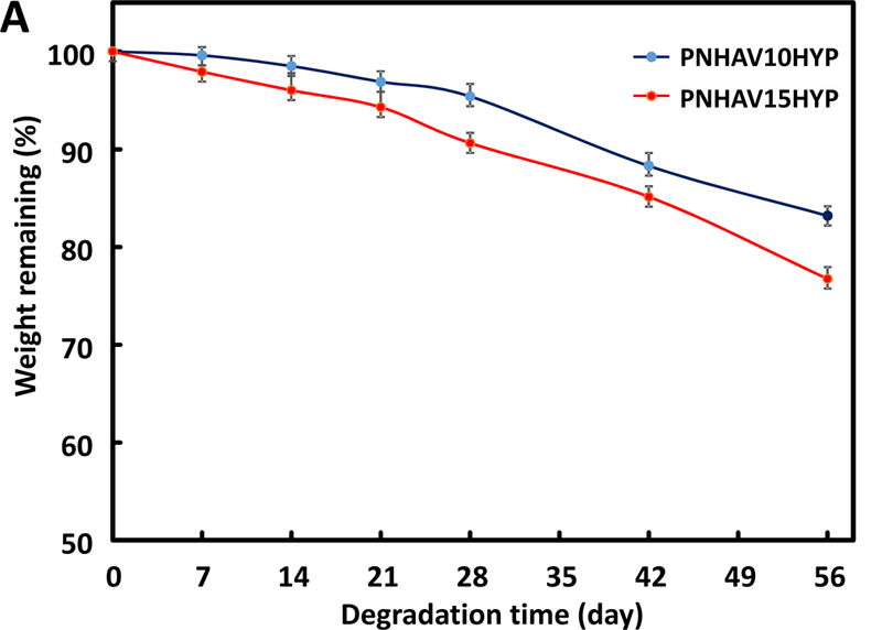
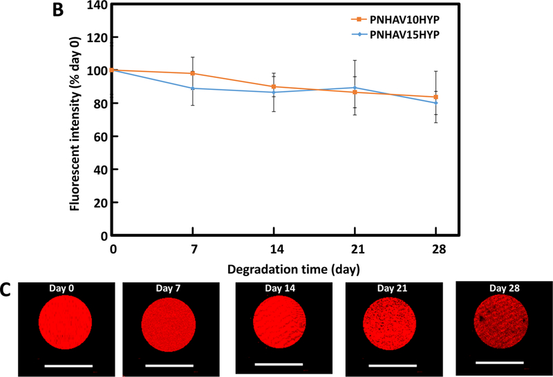
Figure 4.


Hydrogel degradation and fluorescence signal change. (A) weight remaining of hydrogels when incubating in PBS at 37°C for 8 weeks;hydrogel fluorescent intensity change in PBS at 37°C for 4 weeks; and (C) hydrogel fluorescent images during degradation. PNHAV10HYP and PNHAV15HYP represent HYP‐conjugated hydrogels PNHAV with VP content of 10% and 15%, respectively.
