Figure 6.


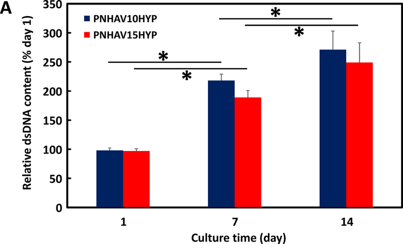
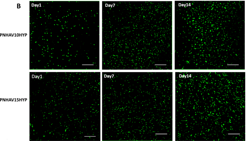
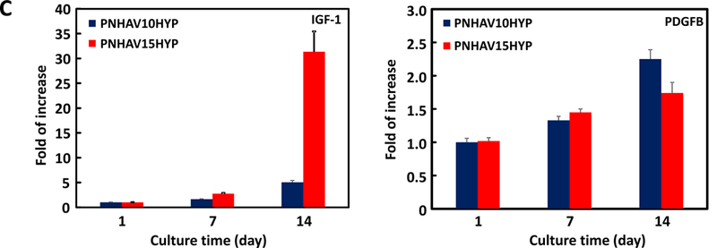
MSC growth in HYP‐conjugated hydrogels. (A) dsDNA content of MSCs encapsulated in the hydrogels during 14 days of culture. * p<0.05; (B) live cell images of MSCs encapsulated in the hydrogels during 14 days of culture period. PNHAV10HYP and PNHAV15HYP represent HYP‐conjugated hydrogels PNHAV with VP content of 10% and 15%, respectively. Scale bar = 200 μm; (C) Paracrine effects of MSCs encapsulated in HYP‐conjugated hydrogels during 14 days of culture. Gene expressions of IGF‐1 and PDGFB were determined by real time RT‐PCR.
