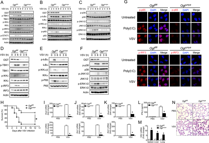
Figure 3.
OGT is critical for the activation of antiviral immune signaling and antiviral innate immune responses in vivo. (A to C) Phosphorylation of IFN-I signaling molecules including TBK1, IKKε, IRF3 and STAT1 (A), NF-κB (B), and MAPK (C) signaling molecules in Ogtfl/fl and OgtΔmye BMMs left untreated or stimulated with VSV (MOI = 1) for indicated periods. (D to F) Phosphorylated TBK1, IKKε, IRF3 and STAT1 (D), NF-κB (E), and MAPK (F) signaling molecules in Ogtfl/fl and OgtΔmye peritoneal macrophages treated with VSV for indicated periods. (G) Immunofluorescence staining of phosphorylated IRF3 (upper panel) or p65 (lower panel) in Ogtfl/fl and OgtΔmye BMMs left untreated or challenged with VSV for 4 h. Scale bar, 20 μm. (H) Survival of Ogtfl/fl (n = 18) and OgtΔmye (n = 20) mice after intraperitoneal injection with VSV (1 × 10⁸ PFU per mouse). (I to L) Transcripts of Ifna4 and Ifnb1 in the spleen (I), liver (J), or lungs (K) and IFN-β protein in serum (L) from Ogtfl/fl and OgtΔmye mice challenged with either PBS or VSV (2 × 10⁷ PFU) for 24 h (n = 6 per group). (M and N) VSV RNA in the spleen, liver and lungs (M) and histological analysis of the lung tissue (N) in Ogtfl/fl and OgtΔmye mice challenged with VSV (n = 5 per group). Scale bar: 20 μm. * P < 0.05, versus controls (Kaplan-Meier (H) or two-tailed Student’s t-test (A to F, I to M)). Data are from one experiment representative of three independent experiments and are expressed as mean ± s.d.

