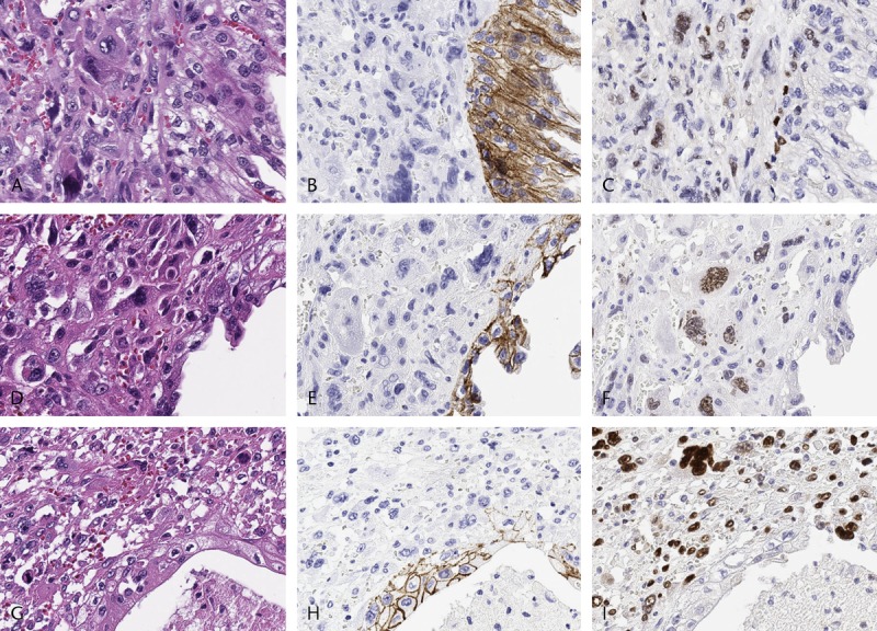
FIGURE 2.

Representative histologic findings and immunoreactivity of E-cadherin and EMT transcription factors in the transitional component of APCs (A–I, original magnification ×400). A–C, D–F, and G–I were observed in the same location. Immunoreactivity of E-cadherin on the cell membrane (B, E, and H). Nuclear expression of Slug (C), Twist (F), and Zeb1 (I) was observed in the transitional component of APCs.
