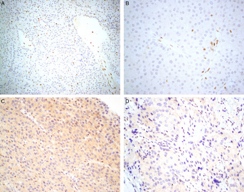
FIGURE 1.

We emphasize that parafibromin can be a difficult stain to perform and interpret and often different conditions are required to achieve a workable result. This figure shows parafibromin IHC from the same tumor performed on the same block initially at primary diagnosis (A, B) and repeated 8 years later for this study (C, D). When first performed, all non-neoplastic cells are completely negative with crisp nuclear staining in internal positive controls (A, B). C, When repeated on archived material, a greater concentration of primary antibody was required to achieve expression in internal positive controls resulting in nonspecific cytoplasmic staining but still completely absent nuclear staining in neoplastic cells. D, The internal controls are weaker in some areas on repeat staining but still positive. Parafibromin IHC, original magnifications A) 200x, B) and C) 400x, D) 600x.
