
FIGURE 6.
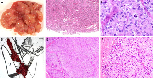
Patient 5’s case was initially classified as adenoma at first excision (A–C) but subsequently demonstrated unequivocal metastasis warranting classification as carcinoma (D–F). At initial resection, the tumor came out easily but was fragmented (A) making assessment of the interface with non-neoplastic tissue difficult. B, However, no unequivocal invasive growth was evident. C, At high power the primary tumor demonstrated cytologic features of parafibromin deficiency, including nuclear enlargement with relatively preserved N/C ratios and eosinophilic but not oxyphilic cytoplasm. D, At recurrence, the tumor was resected from the level 3/4 lymph nodes, effectively excluding seeding of benign disease from the previous operation. The recurrence demonstrated an unequivocal invasive growth pattern into soft tissue (E), but it still showed similar cytologic features to that seen in the original tumor (F). Hematoxylin and eosins, original magnifications. B) 100x, C) 400x, E) 100x , F) 200x.