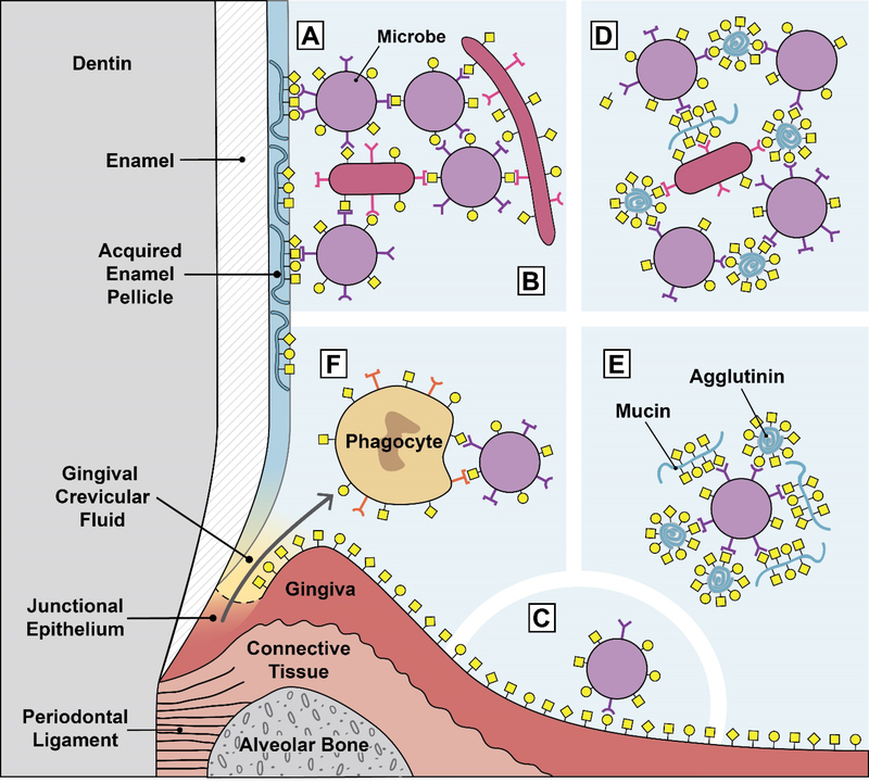
Fig. 1.
Glycan-mediated interactions in the oral environment. A. Microbial attachment to the tooth surface. Microbial adhesins attach to mostly salivary glycoproteins adsorbed to the tooth surface as part of the acquired enamel pellicle. This is typically the first step in oral biofilm formation. B. Microbial adhesins attach to glycans on other microbes. This example shows microbial coadhesion, the process of attaching to microbes that are part of a biofilm. If this microbe-microbe attachment occurs in suspension, i.e. in the planktonic phase, it is called microbial coaggregation. C. Microbial adhesins attach to glycans on oral epithelia. This interaction normally does not result in long-term colonization because the oral epithelium is a shedding surface. D. Salivary glycoproteins agglutinate microbes. Natural salivary flow causes these aggregates to be swallowed, resulting in clearance of the microbes from the oral cavity. E. Salivary glycoproteins can serve as molecular camouflage. A microbial cells is covered by multiple bound salivary agglutinins and mucins. This might protect the microbe from immune surveillance by masking the underlying microbial cell surface. F. Lectin-mediated phagocytosis. Phagocyte lectins bind to microbial glycans, and microbial adhesins bind to phagocyte glycans. These interactions can both potentially lead to phagocyte activation and phagocytosis of the bacteria.

