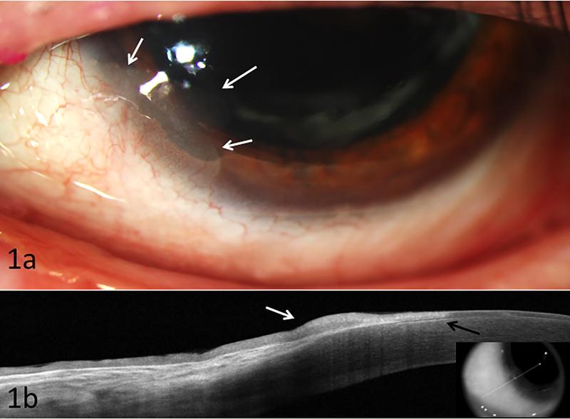
Figure 1:
Biomicroscopy of case 1 illustrating (a) recurrent inferonasal OSSN at 7 o’clock (white arrows) with subepithelial scarring; (b) High resolution ocular coherence tomography demonstrating thickened hyper-reflective epithelium (white arrow) with an abrupt transition from normal to abnormal epithelium (black arrow), consistent with ocular surface squamous neoplasia (OSSN). Note subepithelial scaring under the abnormal epithelium. Biopsy confirmed findings.

