Figure 3:
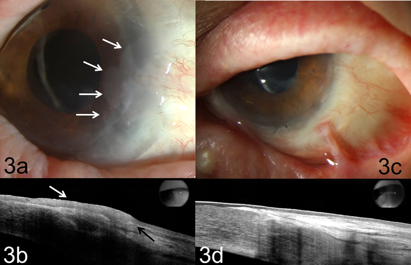
Biomicroscopy of case 3 illustrating (a) a biopsy proven recurrent OSSN with an opalescent and gelatinous lesion at 2 to 4:30 o’clock limbus. There was extension onto corneal surface about 4 mm (white arrows) at edge of the limbal autograft; (b) high resolution OCT (HR-OCT) confirmed recurrent OSSN with thickened, hyper-reflective epithelium (white arrow) and an abrupt transition from normal to abnormal epithelium (black arrow). (c) Tumor resolved OSSN after 4 rounds of 5-FU and retinoic acid; note eyelid scarring from atopic dermatitis. (d) HR-OCT confirms thin, normalized epithelium at resolution of treatment.

