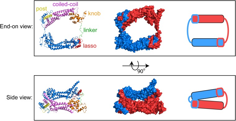
Fig. 2.
Atomic structure of the dimeric FH2 domain of Bni1p. End-on and side views of the dimeric FH2 domain of the S. cerevisiae formin Bni1p. The structure depicts the actin-bound FH2 dimer, based on pdb ID 1Y64 (Otomo et al. 2005), following 160 ns of all-atom molecular dynamics simulations (Baker et al. 2015). Left = Ribbon representations of the FH2 dimer. One monomer is color-coded to highlight the lasso, linker, knob, coiled-coil and post subdomains. The second monomer is depicted in blue. Center = surface representations of the FH2 dimer; the monomers are depicted in red and blue. Right = cartoon representations of the FH2 dimer

