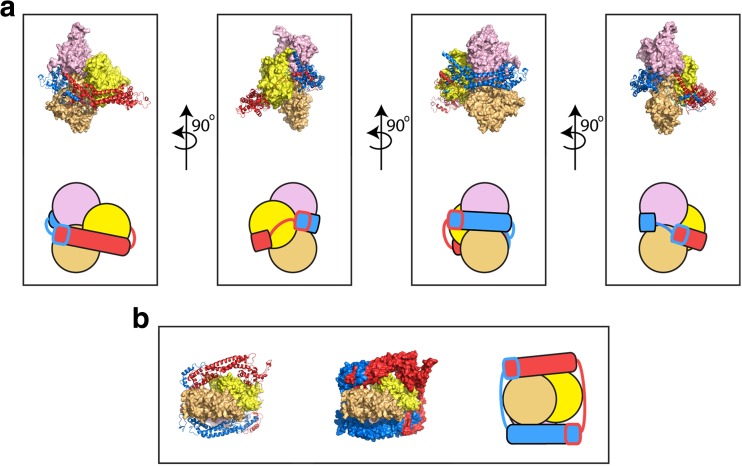
Fig. 3.
Structure of the dimeric FH2 domain of Bni1p bound to actin. Structural representations of the FH2 dimer of the S. cerevisiae formin Bni1p bound to the three terminal subunits of an actin filament following 160 ns of all-atom molecular dynamics simulations (Baker et al. 2015). The initial structural model was constructed based on the crystal structure of the actin-bound Bni1p FH2 domain (pdb ID 1Y64) (Otomo et al. 2005). The structures of the FH2 domains are represented as ribbon diagrams and the individual domains are colored red and blue. The structures of the three actin subunits are depicted as surface representations. The terminal, barbed end subunit is depicted in light orange. The other two actins are shown in yellow and pink. a Multiple orientations of the side view are depicted. The FH2 dimer forms contacts with all three actin subunits. Cartoon representations are also shown to orient the reader. b End-on view of the FH2-actin complex. The FH2 dimer is shown both with ribbon diagrams (right) and as a surface representation (center). A cartoon representation is also shown (left)

