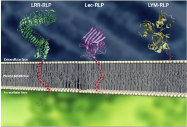
Figure 1: Conceptual representations of RLPs with different ectodomains on the plasma membrane.
From left: LRR-RLP (green), Lec-RLP (purple) and LYM-RLP (gold); transmembrane helices are depicted in red. The transmembrane helices were added to depict a representation of the structure, but do not represent the actual structure of the transmembrane domains of these proteins. The protein structure of OsCEBiP (PDB: 5JCD) represents the LYM in this figure, anchored to the membrane with a GPI anchor. The intracellular area is further indicated by the depiction of short cytoplasmic tails (red). Molecular structures were visualized using Visual Molecular Dynamics (VMD) software [101]. PM and GPI anchor were modeled using Autodesk 3DS Max (2017). Proteins containing domains with similar structure were used to represent RLPs: LRR-RLP (BRI1; PDB: 3RGX4), LecRLP (PHA-E; PDB: 3WCR5) [102, 103].

