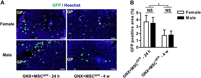
Fig. 5. Bone marrow tracing of infused mesenchymal stem cells (MSCs).
a Immunofluorescent staining of inhabited MSCs in recipient mouse bone marrow of distal femora after systemic transplantation. Donor MSCs were labeled with green fluorescent protein (MSCGFP). Bars: 100 μm. GP growth plate, GNX gonadectomy. b Corresponding quantification of engrafted GFP-labeled MSCs. n = 3 per group. The data represent the means ± SD. *P < 0.05; NS not significant (P > 0.05)

