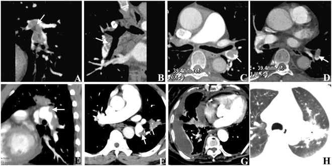
Figure 2.
CT manifestations of pulmonary embolism. (A,B) The sagittal (A) and coronal (B) MPR images showed the right inferior pulmonary artery were filled with a filling defect, which belonged to a complete occlusion type of pulmonary embolism. (C,D) The original transverse axis images showed the central filling defect surrounded by contrast media in the left inferior pulmonary artery, which belonged to a central type of pulmonary embolism. (E,F) The coronal (E) and axis images (F) showed the filling defects in one side of the artery, which belonged to a peripheral type. (G) The pleural effusion and pericardial effusion was showed in one patient. (H) The pulmonary infarction was showed in one patient. Arrows indicate the embolus.

