Fig. 4.
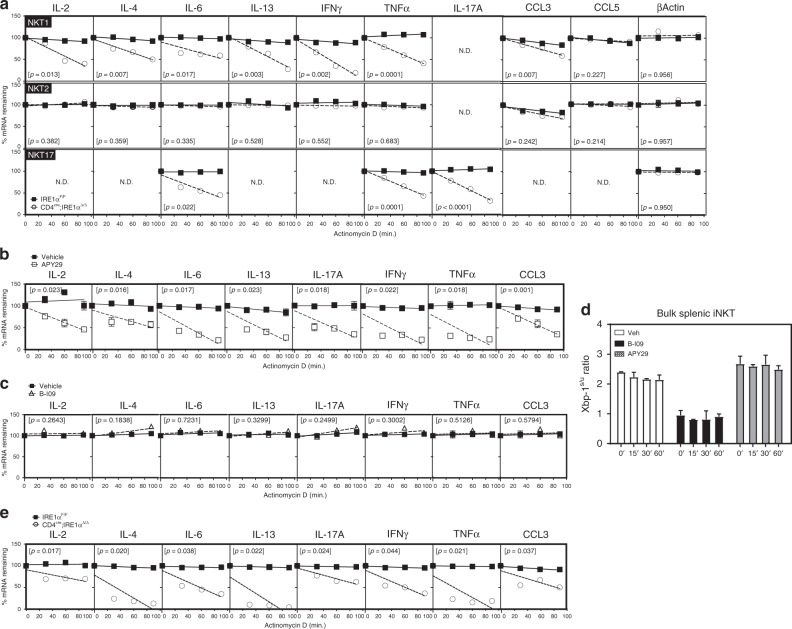
Reduced cytokine mRNA stability in IRE1α-deficient iNKT cells. a Splenic NKT1, 2 and 17 cells were isolated from control (n = 4) and CD4cre;IRE1αΔ/Δ (n = 4) mice, expanded and restimulated with anti-CD3/CD28 for 3 h prior to addition of actinomycin D. mRNA decay rates were determined by linear regression analysis. Data represent three independent experiments pooled. b−d Bulk splenic wild-type iNKT cells were expanded in vitro, restimulated with anti-CD3/CD28 for 3 h in the presence of APY29 and B-109, and then treated with actinomycin D. mRNA decay rates were determined as above. Data represent two independent experiments pooled. e Purified bulk splenic iNKT cells from control (n = 6) and CD4cre;IRE1αΔ/Δ (n = 6) mice were restimulated ex vivo with α-CD3/CD28 for 3 h, prior to addition of actinomycin D. Data represent two independent experiments pooled. Error bars show the mean ± s.e.m. p < 0.05 determined by linear regression analysis and ANOVA, n.d. not detected

