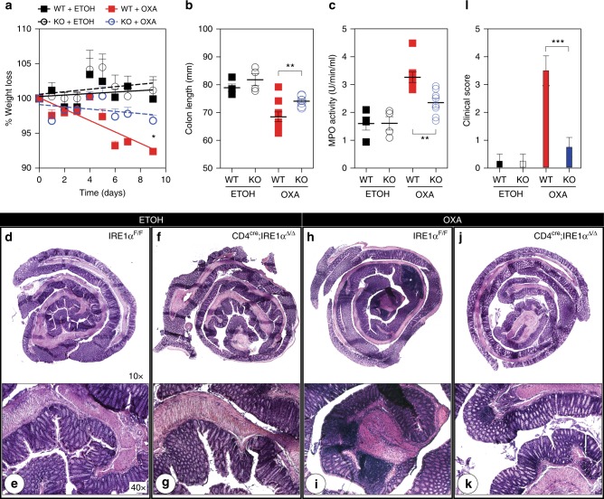
Fig. 6.
Deletion of IRE1α within iNKT cells protects mice from oxazolone colitis. IRE1αF/F control (n = 10) and CD4cre;IRE1αΔ/Δ (n = 10) mice were presensitized with either ethanol (ETOH) or 3% oxazolone (OXA) and then challenged intrarectally with either ethanol or 1% oxazolone 7 days after intrarectal challenge. Weight loss (a), colon length (b), MPO activity (c) and clinical score (l) were subsequently evaluated for each group after the intrarectal challenge. Data represent mean values obtained from two independent experiments. d, f, h, j Photomicrographs (×10) of hematoxylin and eosin (H&E)-stained sections of whole intestine removed from ethanol or oxazolone-treated control (d, h) and CD4cre;IRE1αΔ/Δ (f, j) mice 5 days after intrarectal challenge. e, g, i, k Photomicrographs (×40) represent boxed distal regions of 10× H&E-stained sections representative for each group. Asterisk indicates areas of mucosal erosion. Error bars show the mean ± s.e.m. Mixed model analysis was performed to assess differences in weight loss and *p < 0.05, **p < 0.01, ***p < 0.001 determined by Mann–Whitney U test

