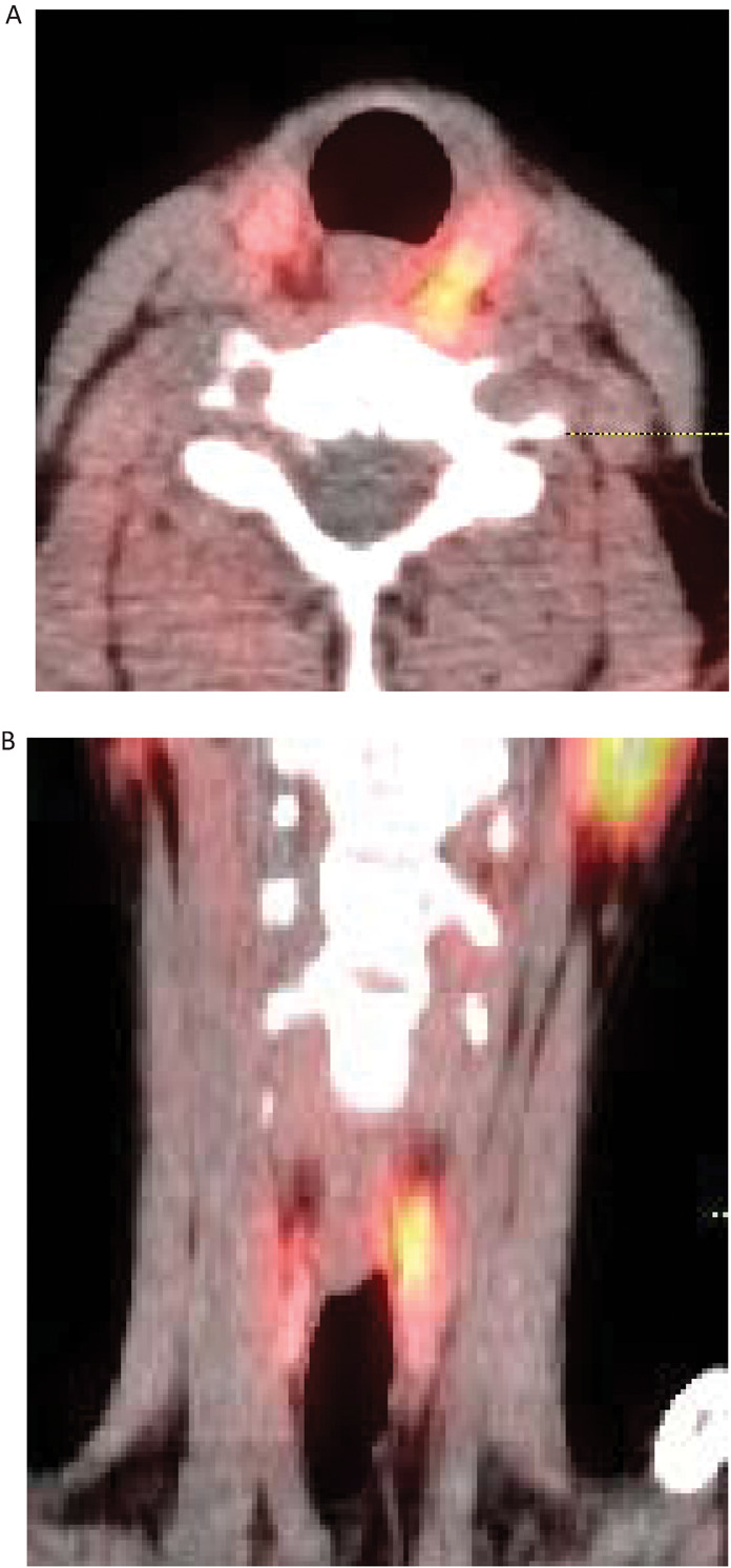
Fig 1.

SPECT CT images of parathyroid adenoma. Fused (A) axial and (B) coronal SPECT CT images showing a superior left parathyroid adenoma adjacent to the oesophagus posterior to the left lobe of thyroid. Physiological uptake in the parotid glands is also seen. SPECT = single photon emission computerised tomography
